Restriction Enzymes: A History
By Wil A.M. Loenen, Leiden University Medical Center
April 2019 · 346 pages, illustrated (38 color and 26 B&W)
ISBN 978-1-621821-05-2
Chapter 2
Chapter doi:10.1101/restrictionenzymes_2
Host-Controlled Variation Is Methylation and Restriction of DNA: The 1960s
INTRODUCTION
Werner Arber entered the restriction field by chance. His research into host-controlled variation (HCV) stands as one of the examples of “serendipity” in scientific discovery: the combination of a chance observation, an opportunity that favors the prepared mind, and being at the right place at the right time. Serendipity has been referred to by Salvador Luria in his autobiography entitled A Slot Machine, a Broken Test Tube (Luria 1984). The slot machine refers to his landmark paper on spontaneous mutation in bacteria with Max Delbrück from 1943 (Luria and Delbruck 1943), the broken test tube to the event that caused him to discover restriction of one of his mutant T* phages: He dropped his bacterial culture and asked Joe Bertani for some other cells. Bertani gave him some Shigella, which happily plated T*, in contrast to the Escherichia coli B/4o cells that had originally produced that phage. (Later it would become clear that T* had become sensitive to the mcr restriction system [see Chapter 8] present in the E. coli B strain, but absent in Shigella.) Arber's chance observation was that the generation of lambdas derivatives of E. coli B/r resulted in strains that did not properly propagate lambda phage, as discussed below.
The Arber–Dussoix Papers (1962)
Werner Arber entered biophysics as a student of Edward Kellenberger in Geneva (Fig. 1) in 1953, the year of the double helix model, the Bertani and Weigle paper, and the first review by Salvador Luria on HCV (Chapter 1). During this period, Arber held a journal club seminar on the Watson and Crick model of the DNA double helix (Watson and Crick 1953). Sixty years later, he recalled that there was very little acceptance that genetic information was present in the linear sequence of base pairs (bp), because people had the idea that genetic information was very complicated. They simply could not understand how nucleic acid, composed of only four different building blocks, the nucleotides (nt), was able to give that information rather than the proteins with 20 building blocks, the amino acids (aa), that would be much better carriers of highly complex information (http://library.cshl.edu/Meetings/restriction-enzymes/v-Arber.php).
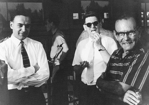
FIGURE 1. (Left to right) Werner Arber, Edouard Kellenberger, and Jean Weigle after the defense of Arber's PhD thesis (1958). (Courtesy of Werner Arber.)
This is in contrast to Matthew (Matt) Meselson's recollection of this event (Meselson with Franklin Stahl provided the experimental evidence for the semiconservative nature of DNA replication in 1958 [Meselson and Stahl 1958]). Meselson recalls that “e.g., Erwin Chargaff (who determined that the ratios of adenine to thymine and of guanine to cytosine were always unity [see Judson 1979 for details]), upon reading the Avery et al. paper of 1944 [Avery et al. 1944] almost immediately stopped his research on lipids and began his well-known work on the nucleotide composition of DNA, from diverse organisms, showing wide variation in composition and thereby overthrowing Levene's tetranucleotide hypothesis. Further work in the late 1940s by Hotchkiss and others [for an overview see, e.g., Hotchkiss 1953, 1995] showed that highly purified DNA could transform specific bacterial genes. The 1952 finding by Al Hershey and Martha Chase [Hershey and Chase 1952] that upon infection most of the 32P of phage T2 enters the bacterial cell while most of the 35S stays outside, together with the earlier transformation experiments, was widely interpreted at the time to mean that the information resides in DNA, not protein” (M Meselson, pers. comm.; references added by the author). There is an interesting anecdote in this respect: Apparently, as late as 1956, Beadle asked an “eminent biochemist” whether he thought the double helix model was correct. The answer: “Yes, I am sure it is correct, but I do not think it has anything to do with replication!” (Berends 1977). But according to Meselson, this was an oddity by then.
This journal club activity and his training in a high-powered laboratory generated Arber's lifelong interest in research into DNA and phage genetics. For his PhD thesis he studied a defective phage called lambda dgal, obtained from Larry Morse in Joshua Lederberg's laboratory (for details of this phage, see http://www.sci.sdsu.edu/∼smaloy/MicrobialGenetics/topics/phage/lambda-dgal.html). Afterward he spent a year with Joe and Betty Bertani to work on P1-mediated transduction of lambda prophage as well as the fertility (F) factor. Here he heard about the 1953 HCV paper. At that time people had started to realize the huge size of genomes of many organisms. How could you study the structure or function of a gene in such a large genome? Impossible! You had to take the gene out. But how? Could you perhaps incorporate genes of interest in a phage? In that case, one could harvest enough DNA for biochemical studies.
At the suggestion of Esther Lederberg, Arber went to work with the radiationr E. coli B/r strain after his return to Geneva. He first made a lambdas derivative of this strain, as E. coli B was not a normal host for lambda (it was used for T phages). However, the new strains did not efficiently propagate lambda (which had been prepared in its normal E. coli K12 host). Fortunately, he quickly realized that this was owing to the HCV phenomenon he had heard about before. Intriguing questions were to be answered: How could phage overcome this barrier? What exactly happened to the phage, when it was prevented to grow normally? How did phage become adapted to the new host? Was HCV caused by a host protein picked up by the phage during its replication that would allow it to return to the same host? Or, alternatively, rather than taking a protein along in the phage head, would the phage DNA get some kind of “imprinting” by the host? Using phage DNA labeled with 32P, he could resolve this issue and test whether the new DNA of the progeny phage had no host modification. Not a protein in the phage head but something on the DNA caused modification.
In the meantime, Grete Kellenberger in the laboratory had found that irradiated phage after infection was rapidly destroyed. She suggested to Arber's PhD student Daisy Dussoix (Fig. 2) that Dussoix should test her restricting strains in the same way. The results of these experiments led to the two classical “Arber–Dussoix” papers on the inactivation of DNA by restriction, and modification of DNA owing to a host function (Arber and Dussoix 1962; Dussoix and Arber 1962). In paper I (Arber and Dussoix 1962) they analyzed the fate of lambda DNA in E. coli K12 carrying one of Bertani's original temperate phages, P1 (Table 1). This strain carries two restriction systems, that of E. coli K12 and phage P1 (“K” and “P1,” currently called EcoKI and EcoP1I, respectively, later designated as members of the Type I and III REases [http://rebase.neb.com/rebase/rebase.html; Roberts et al. 2003, 2015]). Lambda cultured on E. coli C (lacking a restriction system), lambda·C, was restricted cumulatively by both K and P1, leading to a drop in efficiency of plating from 2 × 10⁻⁴ to 7 × 10⁻⁷.
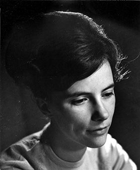
FIGURE 2. Daisy Roulland-Dussoix (∼1962) (Courtesy of André Dussoix.)
|
TABLE 1. Efficiency of plating of phage λ variants on different host strains |
||||
Efficiency of plating on host strains |
||||
Phage variant |
K12 |
K12(P1) |
B251 |
C |
λ · K |
1 |
2 × 10⁻⁵ |
10−4 |
1 |
λ · K(P1) |
1 |
1 |
10−4 |
1 |
λ · B |
4 × 10⁻⁴ |
7 × 10⁻⁷ |
1 |
1 |
λ · C |
4 × 10⁻⁴ |
4 × 10⁻⁷ |
2 × 10⁻⁴ |
1 |
|
Reprinted from Arber and Dussoix 1962, with permission from Elsevier. |
||||
After adsorption of phage onto P1-restricting or nonrestricting cells, the DNA apparently entered both cell types within minutes, as judged from the presence of the label in the pellet after low-speed centrifugation. It became clear that the phage DNA was injected and degraded upon infection of different bacterial hosts (Dussoix and Arber 1962; Lederberg and Meselson 1964) unless it carried host-specific modification of that DNA. Similar DNA breakdown with other restriction systems supported these data.
Grete Kellenberger had already suggested that this degradation might be a two-step process. A highly specific “restriction enzyme” (REase) would be cleaving the DNA, followed by one or more less-specific nuclease(s) that would degrade the cleavage products, a prediction later shown to be correct. Further evidence that restriction occurred at the level of DNA came from the observation that hybrid DNA molecules, with one strand modified and the other not, were not degraded and on transformation gave rise to new phage progeny (Arber and Dussoix 1962; Arber 1965; Meselson and Yuan 1968).
In paper II (Dussoix and Arber 1962), the “suicide” method was used to analyze the nature of HCV of newly synthesized DNA. This protocol was developed by Alfred Hershey in 1951 and extensively used by Gunther Stent: DNA heavily labeled with 32P becomes noninfectious owing to radioactive damage, but the DNA will not be immediately degraded. Thus the modification status of new phage DNA, made in the cell after infection in medium free of label, could be easily analyzed. These experiments proved the modification to be physically linked to the DNA. In this paper, they also provided the first tentative evidence that modification might occur on nonreplicating DNA, and that modification was dominant—that is, the presence of modification on one of the two strands would protect the DNA, as was later shown definitively by the inability of the EcoKI enzyme to make even single-stranded (ss) breaks on lambda DNA having one modified chain and one nonmodified chain (Meselson and Yuan 1968).
The molecular aspects of restriction-modification (R-M) were becoming clear. Both R and M appeared to act somehow on the DNA, and apparently the modification was reversible upon replication, during which it did not lead to mutations. Phage DNA was modified upon its propagation and successfully infected the same host again. In contrast, not properly modified phage DNA was broken down to oligonucleotides in an r+ host. Probably the R-M enzymes could detect minor changes in different DNA molecules. It also became clear that R and M acted on both phage and cellular DNA, a fact we now take for granted. Similar DNA breakdown was observed soon afterward for other restriction systems. In r− mutants the DNA remained intact as predicted.
The three main topics of subsequent study were the nature of the DNA modification, the localization of the host genes for R-M, and the purification of the enzymes responsible for these activities. The Geneva group headed by Arber continued their research resulting in a series of papers during the 1960s. The Americans Stuart (Stu) Linn and William (Bill) Wood came to help with the isolation of the enzymes and mutants, respectively. Stu Linn did his first degree at Caltech and his PhD at Stanford, where he shared a laboratory with Daisy Dussoix. He was a postdoc with Arber from 1966 to 1968 before moving to Berkeley. Bill Wood was a PhD student of Paul Berg, went to Geneva in 1963, and became assistant professor at Caltech afterward. Clearly, R and M were two separate events, and both topics generated widespread interest, inspiring others to embark on the genetics and enzymology of R-M systems.
THE ROLE OF METHIONINE IN DNA MODIFICATION
The first major problem to tackle was the nature of the modification. Although the modification was apparently attached to the DNA in a “phenol-insensitive way” (Dussoix and Arber 1965), there was no evidence about the nature of the actual change. Gunther Stent suggested that perhaps modification was methylation. When Arber visited Berkeley in 1963, he got the first inkling for the essential role of methionine in the modification process: lambda became poorly modified on propagation in an E. coli met− host and withdrawal of this amino acid (aa) from the medium. The essential role for methionine led to the idea of nucleotide “alkylation” of special (presumably sequence-specific) sites on the DNA, via methyl transfer using S-adenosylmethionine (SAM) as donor.
Next, John Smith from Cambridge came to Geneva to look at the correlation of modification with DNA methylation. Unfortunately, basal levels of methyl groups on the lambda DNA were high. Hence, no significant differences in methylation could be found between phage grown on m+ or m− bacteria. This high basal methylation level was not entirely surprising, as the bacterial chromosome was known to be considerably methylated, a property later shown to be due to the action of different bacterial methylases that modify adenine (to m6A) or cytosine (to m5C). The solution to this problem was provided by Hartmut Hoffmann-Berling, who suggested the use of phage fd, which has a single-stranded circular DNA chromosome and is a close relative of the well-known phage M13 of Sanger sequencing fame.
Stu Linn and Werner Arber established that the presence of the E. coli B REase (R·B) led to a drop in the phage fd titer. The phage DNA itself was degraded at a later stage, and in the electron microscope only limited double-stranded (ds) breaks were seen. Phage fd has two recognition sites for the B enzyme (Arber and Kuhnlein 1967), and on the ds-replicative form (RF) of its DNA two As per site were found to be methylated, suggesting one methylated A on each strand (Kuhnlein and Arber 1972). Attempts to determine the nt sequence around this A were initially not successful (van Ormondt et al. 1973; Linn et al. 1974). Phage mutants that lacked these restriction sites were no longer modified. These data proved (1) a link between the number of methylated sites and the restriction targets; (2) the B enzyme methylated an A, presumably within the specificity site; (3) restriction resulted in scission of the DNA, presumably at the same site (which would later prove untrue!); and (4) both activities acted on a substrate lacking this modification. This protective effect to DNA cleavage was the first evidence for a biological function of DNA methylation. In today's terminology this is clearly one of the first described epigenetic effects. In his 1965 review, Arber suggested to name the phenomenon host-controlled modification, as the term variation could indicate some permanent change in the genetic message, which was clearly not the case (Arber 1965). He anticipated that R and M enzymes would be very helpful for functional and structural studies of genetic information.
THE LOCALIZATION AND ALLELISM OF THE GENES ENCODING THE E. coli K12 AND B GENES
The second issue to solve was the location of the R-M genes. Throughout the 1960s, research concentrated mainly on the systems of E. coli K12 and B and phage P1, although restriction activity by so-called “resistance transfer factors” (R or RTF factors) was reported by Tsutomu Watanabe and coworkers in Tokyo during this time. This laboratory was the first to show restriction of infectious lambda DNA in vitro using a bacterial extract containing an R factor, which conferred antibioticr to the host (Takano et al. 1966). Watanabe published groundbreaking papers on this topic (see, e.g., Watanabe 1963; Watanabe et al. 1962, 1964, 1966; Takano et al. 1966, 1968; and Appendix 1 with translation of Japanese Wikipedia entry for Watanabe). This would inspire Herbert (Herb) Boyer to ask PhD student Robert Yoshimori to screen clinical isolates for REases, which led to the discovery of EcoRI (and EcoRII [Yoshimori 1971]; see Chapter 3).
Because of the experimental and genetic tools and knowledge available, the hunt for the genes responsible for R-M was dependent on gene transfer by conjugation and transduction (see Chapters 9 and 13 in Stent and Calendar 1978), before the breakthrough by Mandel and Higa, who managed to transform E. coli with the CaCl2 plus heat shock method in 1970 (Mandel and Higa 1970). Survival and modification of incoming phage or bacterial DNA in the recipient was used to answer questions: Where were the R-M genes on the E. coli chromosome? How many genes were involved? What was the exact nature of the modification? Where was it located? How and when and where was the DNA cut and degraded? Was it the same for the E. coli K12, B, and phage P1 systems? More and more evidence would emerge that the phage P1 system was different from that of E. coli K12 and B, and also from the restriction system that attacked Luria's mutant T* phages, as reported in 1952 (Luria and Human 1952; Chapter 1). These facts slowly emerged and would eventually lead to the current classification in the four types of restriction systems and subdivisions (http://rebase.neb.com/rebase/rebase.html; Roberts et al. 2003, 2015; http://www.library.cshl.edu/Meetings/restriction-enzymes/v-Roberts.php).
The genetic mapping experiments were giving a reasonably coherent picture of the genomic locus encoding the R-M enzymes of E. coli K12 and B. Experiments with partial dipoids (“merozygotes”) provided convincing evidence that (1) K and B mutants (either r−m+ or r−m−) complement each other; (2) the systems were functionally allelic: recombinants with E. coli K12 and B/r restricting and modifying properties never superimposed on one another but were always mutually exclusive (in contrast to the K and P1 activities in the Arber and Dussoix experiments); (3) the R-M genes in both strains had to be linked, as it was easy to transfer them together; and (4) there were three genes located near thr on the E. coli chromosome. The most-conclusive evidence for the three-gene model was obtained with temperature-sensitive (ts) hsm mutants made by Josef Hubáček and Stuart Glover (Hubáček and Glover 1970). All three genes were probably needed for restriction, although the involvement of additional genes could not be formally excluded at the time. The K and B genes were called hss, hsm, and hsr (currently host specificity determinant hsdS, hsdM, and hsdR), and it was tentatively concluded that hsr was not needed for modification. The P1 system would prove to be a two-gene system, named res and mod, respectively.
The picture emerging was quite clear: Modification acted on the DNA itself at a limited number of sites in the shape of methylation, leading to host specificity, and restriction occurred only if proper host specificity was absent. The implications were obvious, and the logical assumption was that both R-M enzymes recognized the same particular base sequences. The enzymes for R and M would thus share that part that recognized these specific sites, so that mutation arising in this part would cause the loss of both functions in question. The same explanation could apply independently of whether only one gene product exerted the functions of sequence recognition, modification, and restriction or whether different gene products were assembled as subunits to the specific enzymes.
The major transition about to occur at the turn of the decade can be easily seen in the reviews published just before and after 1970 (Arber and Linn 1969; Boyer 1971; Meselson et al. 1972; also see Chapter 3). The 1969 review by Arber and Linn discusses three possible models for hyphenated and/or palendromic recognition sites, which are familiar to scientists today. In model I, the specificity would be conferred by one strand only, and the recognition site would be a sequence of nucleotides on that strand with one (perhaps more) modifiable base(s). In model II, both strands would be involved. In this case, a sequence of nucleotide pairs would dictate specificity, with each strand carrying at least one modifiable base. Model III was an extension of the second model in which the specificity site would possess internal symmetry (a palindrome). Such sites could be either contiguous (i.e., all individual bases were essential for recognition to occur) or hyphenated, which would allow one or more ambiguities during recognition. The authors also worked out various estimates of the frequency of the recognition sequence as a function of the length of the specificity site. Based on the number of sites found in phage, this site would be 6- to 8-nt long (later shown to be correct for the enzymes analyzed). Expectations were high that such a putative mechanism of base-sequence specific recognition might provide a tool for the sequence-specific cleavage of DNA. The ability to use different enzymes should allow the sequence determination of DNA molecules. A similar hope (i.e., to use these enzymes to sequence DNA) would lead Richard (Rich) Roberts into the restriction field a few years later. In 1969, the exact sequence of such recognition sites had to await biochemical experiments involving end labeling of broken ends with 32P, and the arrival of DNA sequencing.
In conclusion, during the 1960s, much of the groundbreaking work was performed and many facts became clear that we now so easily take for granted: (1) R and M could take place on nonreplicating DNA; (2) R levels depended on the number of specificity sites per DNA molecule and varied from enzyme to enzyme; and (3) R-M was a general phenomenon: Host, plasmid, and phage DNA were all sensitive to different systems, depending on the bacterial host. Restriction emerged as the bacterial defense system against foreign DNA.
PURIFICATION OF THE RESTRICTION ENZYMES OF E. coli K12 AND B
The groups of Werner Arber in Geneva and Matt Meselson at Harvard University set out to purify the REases from E. coli K12 (EcoKI) and B (EcoBI). Meselson had first detected restriction activity as breakage of unmodified lambda DNA assayed in a sucrose gradient, and started experiments to detect and purify EcoK1. Although he found ATP-dependent EcoKI restriction activity in crude extracts, he could not detect the activity in DEAE column eluates. At that point, Robert (Bob) Yuan came into his laboratory. They used a combination of column chromatography, glycerol gradients, and preparative gels to isolate EcoKI (Meselson and Yuan 1968). They knew that Bill Wood had found evidence that restriction is impaired in methionine auxotrophs if methionine is withheld, so they added methionine and ATP, which restored the activity. But upon further purification the activity again vanished, until they realized that the enzyme needed to be replenished with ATP and that the enzyme also needed SAM for activity (Meselson and Yuan 1968; see Appendix 2 with emails [July 2, 2004] between Mattt Meselson and Noreen Murray for an historical account). Once this absolute requirement for SAM became evident, EcoKI could be purified ∼5000-fold to homogeneity, as determined by gel electrophoresis. During these purification attempts they used breakage of lambda DNA in the presence of ATP, SAM, and Mg2+ as an assay (Meselson and Yuan 1968). For later studies of enzyme binding to DNA and ATP hydrolysis a rather simple and convenient assay was used (Yuan and Meselson 1970; Yuan et al. 1972); filter retention of unmodified DNA. If the EcoKI enzyme was incubated with unmodified and modified lambda DNA and the mixture passed through a nitrocellulose filter, only the unmodified DNA was retained on the filter. Maximum retention required ATP, SAM, and Mg2+, and EcoKI mutant enzymes failed to cause retention.
The enzyme did not break at specific sites and broke the strands sequentially (Meselson and Yuan 1968). The molecular weight (MW) of the native enzyme was ∼400 kDa, based on sedimentation and gel filtration rates relative to proteins of known MW. The complex could be dissociated with SDS, revealing three subunits with MWs of 135, 62, and 52 kDa, respectively. The relative amounts indicated that the complex contained two of each of the larger subunits and only one of the smallest (Meselson et al. 1972). The complex had both REase and MTase activities.
In Geneva, and then in Berkeley, Stu Linn started out to purify EcoBI (Linn and Arber 1968; Eskin and Linn 1972a,b). The protocol was a little different, and eventually yielded a ∼1000-fold purification. Around the same time, Daisy Roulland-Dussoix also purified EcoBI ∼1000-fold in the laboratory of Herb Boyer (Roulland-Dussoix and Boyer 1969). The enzyme proved to be very similar to EcoKI: a large complex with a MW of ∼400 kDa and three types of subunits of MW 135, 60, and 55 kDa (Arber and Linn 1969; Boyer 1971; Eskin and Linn 1972b). It also had an absolute requirement for SAM, needed Mg2+ and ATP as cofactors, and degraded ATP during the reaction. However, in contrast with the stable pentameric EcoKI enzyme, EcoBI purified as several active oligomeric species with a MW ranging from 450 to 750 kDa. Was this difference due to differences in the purification procedures? The predominant form had a proposed subunit composition of two larger, four medium, and two of the smallest subunits (Eskin and Linn 1972b). Two active forms, which were enzymatically indistinguishable, were also isolated by native gel electrophoresis. What was the role of SAM in the restriction process? Technical reasons (the small number of enzyme molecules in the cell and the instability of SAM) made it hard to address this issue (Linn et al. 1977). And why did the enzymes remain attached to the DNA after restriction? Was this a signal to another enzyme molecule (Rosamond et al. 1979)?
The EcoBI MTase, M·EcoBI, could be purified by essentially the same procedure used to prepare the EcoBI REase except that the MTase was separated from the REase during column chromatography (Kuhnlein et al. 1969) and further purified on DNA cellulose (Lautenberger and Linn 1972). M·EcoBI contained the same small subunits as the EcoBI REase (Lautenberger and Linn 1972). Depending on the time after purification, the enzyme had various subunit compositions upon storage, all enzymatically active (Linn et al. 1974). An attempt to match the enzyme subunits to the respective genes proved difficult. Bacteria with mutations in the three hsd genes could complement in vivo, as mentioned earlier, but also in vitro (Linn and Arber 1968). Also, purified MTase could supply the two subunits to generate restriction in hsdS− and hsdM− extracts (Linn 1974). This indicated that the M and S subunits of the MTase had an active role in restriction. In this way, it could be established that the large subunit of 130 kDa was the subunit encoded by the hsdR gene. At the time, the two smaller subunits could not be assigned to either the hsdM or hsdS genes (Linn 1974).
For further details on this topic, see Arber et al. (1975) and Endlich and Linn (1981); for a biography of Daisy Roulland-Dussoix, see Appendix 3; and for more background reading, see Hershey (1971) and Hayes (1968), Stahl (1969), Watson (1970), Portugal and Cohen (1977), Gribbin (1985), Cairns et al. (1992), and Holmes (2001).
REFERENCES
Arber W. 1965. Host-controlled modification of bacteriophage. Ann Rev Microbiol 19: 365–378. 10.1146/annurev.mi.19.100165.002053
Arber W, Dussoix D. 1962. Host specificity of DNA produced by Escherichia coli. I. Host controlled modification of bacteriophage lambda. J Mol Biol 5: 18–36. 10.1016/S0022-2836(62)80058-8
Arber W, Kuhnlein U. 1967. [Mutational loss of the B-specific restriction in bacteriophage fd]. Pathol Microbiol 30: 946–952.
Arber W, Linn S. 1969. DNA modification and restriction. Ann Rev Biochem 38: 467–500. 10.1146/annurev.bi.38.070169.002343
Arber W, Yuan R, Bickle TA. 1975. Strain-specific modification and restriction of DNA in bacteria. In Proceedings of the FEBS Ninth Meeting, Budapest 1974. Post-synthetic modification of macromolecules (ed. Antoni F, Farago A), Vol. 34, pp. 3–22. FEBS, Oxford.
Avery OT, Macleod CM, McCarty M. 1944. Studies on the chemical nature of the substance inducing transformation of pneumococcal types: induction of transformation by a desoxyribonucleic acid fraction isolated from pneumococcus type Iii. J Exp Med 79: 137–158. 10.1084/jem.79.2.137
Berends W. 1977. ‘Biochemistry Lecture Notes,’ Leiden University, The Netherlands. Translation by the author: p. 323.
Boyer HW. 1971. DNA restriction and modification mechanisms in bacteria. Ann Rev Microbiol 25: 153–176. 10.1146/annurev.mi.25.100171.001101
Cairns J, Stent GS, Watson JD. 1992. Phage and the origins of molecular biology, expanded ed. Cold Spring Harbor Laboratory Press, Cold Spring Harbor, NY.
Dussoix D, Arber W. 1962. Host specificity of DNA produced by Escherichia coli. II. Control over acceptance of DNA from infecting phage lambda. J Mol Biol 5: 37–49. 10.1016/S0022-2836(62)80059-X
Dussoix D, Arber W. 1965. Host specificity of DNA produced by Escherichia coli. IV. Host specificity of infectious DNA from bacteriophage lambda. J Mol Biol 11: 238–246. 10.1016/S0022-2836(65)80054-7
Endlich B, Linn S. 1981. Type I restriction enzymes. In The enzymes, 3rd ed. (ed. Boyer PD), Vol XIV, Part A, pp. 137–156. Academic, New York.
Eskin B, Linn S. 1972a. The deoxyribonucleic acid modification and restriction enzymes of Escherichia coli B. J Biol Chem 247: 6192–6196.
Eskin B, Linn S. 1972b. The deoxyribonucleic acid modification and restriction enzymes of Escherichia coli B. II. Purification, subunit structure, and catalytic properties of the restriction endonuclease. J Biol Chem 247: 6183–6191.
Gribbin J. 1985. In search of the double helix. Quantum physics and life. McGraw-Hill, New York.
Hayes W. 1968. The genetics of bacteria and their viruses. Studies in basic genetics and molecular biology, 2nd ed. Blackwell Scientific, Oxford.
Hershey AD. ed. 1971. The bacteriophage lambda. Cold Spring Harbor Laboratory, Cold Spring Harbor, NY.
Hershey AD, Chase M. 1952. Independent functions of viral protein and nucleic acid in growth of bacteriophage. J Gen Physiol 36: 39–56. 10.1085/jgp.36.1.39
Holmes FL. 2001. Meselson, Stahl, and the replication of DNA. A history of the most beautiful experiment in biology. Yale University Press, New Haven, CT.
Hotchkiss RD. 1953. The genetic chemistry of the pneumococcal transformations. Harvey Lect 49: 124–144.
Hotchkiss RD. 1995. DNA in the decade before the double helix. Ann NY Acad Sci 758: 55–73. 10.1111/j.1749-6632.1995.tb24809.x
Hubáček J, Glover SW. 1970. Complementation analysis of temperature-sensitive host specificity mutations in Escherichia coli. J Mol Biol 50: 111–127. 10.1016/0022-2836(70)90108-7
Judson HF. 1979. The eighth day of creation. The makers of the revolution in biology, a Touchstone book. Simon and Schuster, New York.
Kellenberger G, Zichini ML, Weigle JJ. 1961. Exchange of DNA in the recombination of bacteriopage λ. Proc Natl Acad Sci 47: 869–878. 10.1073/pnas.47.6.869
Kuhnlein U, Arber W. 1972. Host specificity of DNA produced by Escherichia coli. XV. The role of nucleotide methylation in in vitro B-specific modification. J Mol Biol 63: 9–19. 10.1016/0022-2836(72)90518-9
Kuhnlein U, Linn S, Arber W. 1969. Host specificity of DNA produced by Escherichia coli. XI. In vitro modification of phage fd replicative form. Proc Natl Acad Sci 63: 556–562. 10.1073/pnas.63.2.556
Lautenberger JA, Linn S. 1972. The deoxyribonucleic acid modification and restriction enzymes of Escherichia coli B. I. Purification, subunit structure, and catalytic properties of the modification methylase. J Biol Chem 247: 6176–6182.
Lederberg S, Meselson M. 1964. Degradation of non-replicating bacteriophage DNA in non-accepting cells. J Mol Biol 8: 623–628. 10.1016/S0022-2836(64)80112-1
Linn S, Arber W. 1968. Host specificity of DNA produced by Escherichia coli, X. In vitro restriction of phage fd replicative form. Proc Natl Acad Sci 59: 1300–1306. 10.1073/pnas.59.4.1300
Linn S, Eskin B, Lautenberger JA, Lackey D, Kimball M. 1977. Host-controlled modification and restriction enzymes of Escherichia coli B and the role of adenosylmethionine. In The biochemistry of adenosylmethionine (ed. Salvatore F, et al.), pp. 521–535. Columbia University Press, New York.
Linn S, Lautenberger B, Eskin B, Lackey D. 1974. The host-controlled restriction and modification enzymes of E. coli B. Fed Proc 33: 1128–1134.
Luria SE. 1984. A slot machine, a broken test tube: an autobiography. Harper & Row, New York.
Luria SE, Delbruck M. 1943. Mutations of bacteria from virus sensitivity to virus resistance. Genetics 28: 491–511.
Luria SE, Human ML. 1952. A nonhereditary, host-induced variation of bacterial viruses. J Bacteriol 64: 557–569.
Mandel M, Higa A. 1970. Calcium-dependent bacteriophage DNA infection. J Mol Biol 53: 159–162. 10.1016/0022-2836(70)90051-3
Meselson M, Stahl FW. 1958. The replication of DNA in Escherichia coli. Proc Natl Acad Sci 44: 671–682. 10.1073/pnas.44.7.671
Meselson M, Yuan R. 1968. DNA restriction enzyme from E. coli. Nature 217: 1110–1114. 10.1038/2171110a0
Meselson M, Yuan R, Heywood J. 1972. Restriction and modification of DNA. Ann Rev Biochem 41: 447–466. 10.1146/annurev.bi.41.070172.002311
Portugal FH, Cohen JS. 1977. A history of the discovery of the structure and function of the genetic substance. MIT (to the Lighthouse Press), Boston.
Roberts RJ, Belfort M, Bestor T, Bhagwat AS, Bickle TA, Bitinaite J, Blumenthal RM, Degtyarev S, Dryden DT, Dybvig K, et al. 2003. A nomenclature for restriction enzymes, DNA methyltransferases, homing endonucleases and their genes. Nucl Acids Res 31: 1805–1812. 10.1093/nar/gkg274
Roberts RJ, Vincze T, Posfai J, Macelis D. 2015. REBASE—a database for DNA restriction and modification: enzymes, genes and genomes. Nucl Acids Res 43: D298–D299. 10.1093/nar/gku1046
Rosamond J, Endlich B, Telander KM, Linn S. 1979. Mechanisms of action of the type-I restriction endonuclease, ecoB, and the recBC DNase from Escherichia coli. Cold Spring Harb Symp Quant Biol 43(Pt 2): 1049–1057. 10.1101/SQB.1979.043.01.114
Roulland-Dussoix D, Boyer HW. 1969. The Escherichia coli B restriction endonuclease. Biochim Biophys Acta 195: 219–229. 10.1016/0005-2787(69)90618-2
Spector DH, Smith K, Padgett T, McCombe P, Roulland-Dussoix D, Moscovici C, Varmus HE, Bishop JM. 1978. Unaffected avian cells contain RNA related to the transforming gene of avian sarcoma viruses. Cell 13: 371–379. 10.1016/0092-8674(78)90205-2
Stahl FW. 1969. The mechanisms of inheritance, 2nd ed. Foundations of modern genetics series (ed. Suskind SS, Hartman PE). Prentice-Hall, Englewood Cliffs, NJ.
Stent GS, Calendar R. 1978. Molecular genetics. An introductory narrative, 2nd ed. W.H. Freeman, San Francisco.
Takano T, Watanabe T, Fukasawa T. 1966. Specific inactivation of infectious lambda DNA by sonicates of restrictive bacteria with R factors. Biochem Biophys Res Commun 25: 192–198. 10.1016/0006-291X(66)90579-1
Takano T, Watanabe T, Fukasawa T. 1968. Mechanism of host-controlled restriction of bacteriophage lambda by R factors in Escherichia coli K12. Virology 34: 290–302. 10.1016/0042-6822(68)90239-0
van Ormondt H, Lautenberger JA, Linn S, de Waard A. 1973. Methylated oligonucleotides derived from bacteriophage fd RF-DNA modified in vitro by E. coli B modification methylase. FEBS Lett 33: 177–180. 10.1016/0014-5793(73)80186-3
Watanabe T. 1963. Infective heredity of multiple drug resistance in bacteria. Bacteriol Rev 27: 87–115.
Watanabe T, Fukasawa T, Takano T. 1962. Conversion of male bacteria of Escherichia coli K12 to resistance to f phages by infection with the episome “resistance transfer factor.” Virology 17: 217–219. 10.1016/0042-6822(62)90108-3
Watanabe T, Nishida H, Ogata C, Arai T, Sato S. 1964. Episome-mediated transfer of drug resistance in enterobacteriaceae. VII. Two types of naturally occurring R factors. J Bacteriol 88: 716–726.
Watanabe T, Takano T, Arai T, Nishida H, Sato S. 1966. Episome-mediated transfer of drug resistance in Enterobacteriaceae. X. Restriction and modification of phages by fi− R factors. J Bacteriol 92: 477–486.
Watson JD. 1970. Molecular biology of the gene, 2nd ed. W.A. Benjamin, New York.
Watson JD, Crick FH. 1953. Molecular structure of nucleic acids: a structure for deoxyribose nucleic acid. Nature 171: 737–738. 10.1038/171737a0
Yoshimori R. 1971. “A genetic and biochemical analysis of the restriction and modification of DNA by resistance transfer factors.” PhD thesis, University of California, San Francisco.
Yoshimori R, Roulland-Dussoix D, Boyer HW. 1972. R factor–controlled restriction and modification of deoxyribonucleic acid: restriction mutants. J Bacteriol 112: 1275–1279.
Yuan R, Heywood J, Meselson M. 1972. ATP hydrolysis by restriction endonuclease from E. coli K. Nature: New Biol 240: 42–43.
Yuan R, Meselson M. 1970. A specific complex between a restriction endonuclease and its DNA substrate. Proc Natl Acad Sci 65: 357–362. 10.1073/pnas.65.2.357
WWW RESOURCES
https://en.wikipedia.org/wiki/Daisy_Roulland-Dussoix A biography of Daisy Roulland-Dussoix.
https://en.wikipedia.org/wiki/Grete_Kellenberger-Gujer A biography of Grete Kellenberger-Gujer.
http://www.estherlederberg.com/home.html The Esther M. Zimmer Lederberg memorial website.
http://library.cshl.edu/Meetings/restriction-enzymes/v-Arber.php History of restriction enzymes meeting archive.
http://www.library.cshl.edu/Meetings/restriction-enzymes/v-Roberts.php History of restriction enzymes archive.
http://rebase.neb.com/rebase/rebase.html A restriction enzyme database.
http://www.sci.sdsu.edu/∼smaloy/MicrobialGenetics/topics/phage/lambda-dgal.html Formation of lambda LFT and HFT lysates.
https://en.wikipedia.org/wiki/Esther_Lederberg A biography of Esther Lederberg.
APPENDIX 1: TSUTOMU WATANABE1
Born in Gifu city, Gifu prefecture. Graduated from Keio Gijuku University School of Medicine in 1948 and studied microbial genetics. He spent his entire life in the study of the mechanism involved in the acquisition of drug resistance by bacteria until his death by stomach cancer on Nov. 4, 1972, and achieved major contributions in not only modern molecular biology but also therapeutic medicine and public health. In the early decades of his scientific life, he carefully examined the genetic mechanism by which bacteria become resistant to streptomycin, a drug that produced much benefit in the treatment of tuberculosis in those days, and proved experimentally the “spontaneous mutation and selection” mechanism. This achievement brought revision to the then-prevailing hypothesis that resistance occurred by “induction” by drugs. The latter half of his scientific life began with the discovery that the simultaneous acquisition of resistance to many drugs by various pathogenic bacteria occurs by the transmission of extranuclear genetic material; he created the name resistance transfer factor (RTF) for this material. Furthermore, he found that RTF belongs to the category of episomes of François Jacob, through examination from various angles. These studies represent a major achievement in modern molecular biology, especially molecular genetics, as attested to by his invitation to a number of international meetings and his giving of memorable lectures on these occasions. For this achievement he received the award from the Japanese Society of Bacteriology and the Purkinje prize from the Czechoslovak Academy of Sciences. It should be emphasized that these studies gave theoretical foundations on the rational use of antibiotics in the treatment of infectious disease patients. That is, if we make a mistake in the proper use of antibiotics, the most powerful weapon for us in the continuing battle between humans and microorganisms, it will cause the extensive spread of resistant microorganisms in nature. He appealed to the scientists, doctors, and public about the dangers of the spreading multidrug-resistant plasmids and warned about the use of excessive amounts of antibiotics for farm animals as well as in aquaculture. In his last years, he tried to examine this situation himself by collaborating with the Egusa laboratory of Tokyo University School of Agriculture, Department of Fishery. He spent most of his scientific life in the Department of Microbiology, Keio University School of Medicine, taught numerous medical students, and trained many research scientists. He also taught molecular biology as a part-time lecturer at Tokyo University School of Agriculture and at Ochanomizu University School of Science.
APPENDIX 2: E-MAILS BETWEEN MATT MESELSON AND NOREEN MURRAY
Re: Your question about EcoKI
Matthew S. Meselson <msm@wjh.harvard.edu> Mon, Jul 5, 2004 at 9:09 PM
To: Noreen Murray <Noreen.Murray@ed.ac.uk>
Cc: ‘PTASHNE, Mark -- Mark Ptashne’ <m-ptashne@mskmail.mskcc.org>, Mark Ptashne <m-ptashne@ski.mskcc.org>, ‘Matthew S. Meselson’ <msm@wjh.harvard.edu>
Dear Noreen,
We always wondered about Jean and Joe's UV effect and had only hand-waving explanations for it. Astounding that, already knowing not to attack semi-methylated DNA, the minuscule bacteria know to inactivate restriction if their own DNA fails to get methylated. Congratulations on your elucidation of the mechanism.
Last October Mark Ptashne asked me about the isolation of EcoKI. So I have taken the easy way out by pasting my reply to him below. After e-mailing it, I found or maybe Mark told me that I had confused John Lis with Stuart Linn, for which I am embarrassed.
You are right about Bill Wood and methionine. Seymour Lederberg and I had earlier done some things with restriction degradation of DNA that made us look into it ourselves. As you are delving into history, you may like to have the list of our restriction-modification publications below, including the one with Seymour.
At the end of what I sent to Mark, I have added an account of the wrong hypothesis about lambda injection that caused me to try ATP at the start of my attempt to isolate a K restriction enzyme. Again taking the easy way out, I am copying this e-mail to Mark.
————————————
Matthew Meselson
Department of Molecular and Cellular Biology
Harvard University
7 Divinity Avenue
Cambridge, MA 02138 USA
email: <msm@wjh.harvard.edu>
telephone: (617) 495-2264
telefax: (617) 496-244
******
‘Luria had our MS for PNAS for many weeks before telling me that I should shorten it. So I sent it to Nature. As was the practice then, I sent a pre-publication copy of the MS to colleagues. That was at about the time we sent it to Salva for PNAS. I sent a copy to John Lis [read Stuart Linn] who was with Arber and maybe a copy to Bill Wood.
The whole trick to isolating the type I restriction enzymes was to know the right co-factors. I had discovered the ATP requirement in a somewhat hilarious way because of a wrong hypothesis about phage lambda injection that you can get me to tell you about over a drink. So long as the lysate was fairly crude, ATP was all that was needed for endonucleolytic action. Adding ATP to crude lysate of restricting bacteria, I got good cleavage of tritium-labeled unmodified lambda DNA with P-32 labeled modified DNA (or the reverse) as control. I measured cleavage by sedimentation in sucrose gradients and dripping and distinguished the radioactive isotopes in Ed Lenhof's scintillation counters on the fifth floor of the Biolabs.
At that point, Bob Yuan came and I invited him to join me in attempting purification of the presumed enzyme. During initial steps of purification the activity went down. But from some experiments of Bill Wood on the effect of methionine on restriction we got the idea that methionine might also be required. With ATP and methionine added we continued to purify but again the activity went down as purification continued. At that point Bob Yuan, knowing biochemistry which he learned in part from Bernie Horecker, and which as you know I do not know very well, realized that S-adenosylmethyltransferase in the still impure preparation might be making SAM from methionine and ATP.
At first, we thought that only SAM would be needed. But it soon turned out that SAM and ATP are both needed, allowing us to purify the endonuclease to “homogeneity”. We discovered to our surprise that even in the limit digest, not all lambda molecules are broken in the same places. The argument followed from the sedimentation distribution of the digested lambda pieces. For example, although there were pieces of size in the range of 40 percent of a lambda chromosome, they accounted for less than 40 mass percent of the digest. (The DNA moves with respect to the enzyme for variable distances from its recognition sites.) This, alas, made the type I endos not useful for genetic engineering. In the same Nature paper we showed that the enzyme nicks before it makes a double-strand break and that hybrid lambda DNA made by annealing modified and unmodified strands is neither nicked nor cleaved, meaning that the enzyme looks at both chains before deciding what to do.
Whether Linn and Arber independently discovered the rather complex co-factor requirements of the endonuclease or instead first learned of the requirements from our MS I do not know.
Looking at Werner's Nobel account, I gather that they got the co-factor requirements for the B restriction endonuclease by assuming they were the same as those we had found for the K enzyme.
******
ABOUT LAMBDA AND ATP: I had been trying entirely without success to find an endonuclease activity in Rec+ coli not present in Rec−. I had isolated some Rec− mutants even before I knew of John Clark's work at Berkeley. For an assay, I looked for reduction in sedimentation velocity of P-32 labeled lambda DNA in sucrose gradients. Finally, dispirited, disgusted and feeling that I had to prove to myself that I was not simply incompetent in biochemistry, I decided to abandon recombination enzymes and look for some other kind of endonuclease--a restriction endonuclease. The assay I had been using for Rec+ endo could be made better by mixing modified and non-modified lambda DNA, one labeled with 32-P and the other with 3-H. Now, there were six tubes in the swinging bucket rotor I had for the experiment. One tube for the mix by itself, one for the mix with r- bacterial extract, one for r+ extract but what to do with the other three tubes? I decided to use up two of the remaining tubes by adding ATP and can't remember what I did with the sixth tube. Why ATP? I knew that cyanide and azide prevent phage T4 injection. Phage workers used azide to synchronize T4 injections. So I thought maybe lambda too needed ATP for injection. The next step in this faulty argument came from asking where would you fight an invader. At the point of entry -- the city gates-- of course. So that is where cells should position their restriction apparatus to defend against hostile DNA. Then it could be that the apparatus that participated in injection and needed ATP to do so would also need ATP to do restriction. A real non-sequitur. Still, it “smelled” right and even though it wasn't right, it worked. From then on, one could try to be a biochemist, as related above.
Of course another reason for adding ATP is that biochemists always add ATP!
******
Ihler, G. and M. Meselson. 1963. Genetic Recombination in Bacteriophage lambda by Breakage and Joining of DNA Molecules. Virology, 21: 7–10.
Lederberg, S. and M. Meselson. 1964. The Degradation of Non-Replicating Bacteriophage DNA in Non-accepting Cells. Journal of Molecular Biology, 8: 623–628.
Menninger, J.R., M. Wright, L. Menninger, and M. Meselson. 1968. Attachment and Detachment of Bacteriophage lambda DNA in Lysogenization and Induction. Journal of Molecular Biology, 32: 631–637.
Meselson, M. and R. Yuan. 1968. An Endonuclease of Host-Controlled Restriction in E. coli. Federation Proceedings, 27: 395.
Meselson, M. and R. Yuan. 1968. DNA Restriction Enzyme from E. coli. Nature, 217: 1110–1114.
Yuan, R. and M. Meselson. 1969. Binding of lambda DNA by a Restriction Endonuclease. Federation Proceedings, 28: 465.
Yuan, R. and M. Meselson. 1970. A Specific Complex Between a Restriction Endonuclease and its DNA Substrate. PNAS, 65: 357–362.
Meselson, M. and R. Yuan. 1971. DNA Restriction Enzyme from E. coli. Procedures in Nucleic Acid Research, eds. G. Cantoni and R. Davies, Harper and Row, New York, 2: 889–895.
Yuan, R. and M. Meselson. 1971. DNA Restriction Enzyme from E. coli. Methods in Enzymolgy, eds. Grossman and Moldave, Academic Press, 21: 269.
Meselson, M., R. Yuan, and J. Heywood. 1972. Restriction and Modification of DNA. Annual Review of Biochemistry, 41: 447–466.
Haberman, A., J. Heywood, and M. Meselson. 1972. DNA Modification Methylase Activity of E. coli Restriction Endonuclease K and P. PNAS, 69: 3138–3141.
Yuan, R., J. Heywood, and M. Meselson 1972. ATP Hydroysis by Restriction Endonuclease from E. coli. K. Nature New Biology, 240: 42–43.
******
>From Arber's Nobel Address:
“This work would not have been possible without a very fruitful help by a large number of collaborators in my own laboratory and of colleagues working on related topics in their own laboratories. I was extremely lucky to receive in my laboratory in the basement of the Physics Institute of the University of Geneva a number of first class graduate students, postdoctoral fellows and senior scientists. It is virtually impossible to list them all in this context, but my warmest collective thanks go to all of them. In 1964 Bill Wood laid out a solid basis for the genetics of the restriction and modification systems EcoK and EcoB. Later, Stuart Linn, profiting from his fruitful contacts with Bob Yuan and Matt Meselson, who worked in the USA on the enzymology of EcoK restriction, set the basis for in vitro studies with EcoB restriction and modification activities. These studies culminated in the final proof that modification in E. coli B and K is brought about by nucleotide methylation. This concept had found its first experimental evidence during my two months' visit in 1963 with Gunther Stent at the University of California in Berkeley. Several years later Urs Kühnlein, a Ph.D. student, and John Smith, working for various lengths of time with us, succeeded in careful in vivo and in vitro measurements on methylation to validate and extend the earlier conclusions. Their experiments also brought important conclusions with regard to the concept of the sites of recognition on the DNA for the restriction and modification enzymes.”
******
On Fri, 2 Jul 2004, Noreen Murray wrote:
Date: Fri, 2 Jul 2004 14:05:42 +0100
From: Noreen Murray <Noreen.Murray@ed.ac.uk>
To: Professor Matthew S Meselson <msm@wjh.harvard.edu>
Subject: History of the purification of EcoKI
Dear Matt,
I write to ask if you would enlighten me of some early facts re: the history of the purification of EcoKI. I have often wondered how you managed to find the appropriate co-factors to permit the purification of this complex enzyme. It is difficult to appreciate how in 1967 it would have been obvious that either ATP or AdoMet (SAM) would be cofactors, although I am aware that the choice of “SAM” was stimulated by the unpublished observation by Bill Wood that restriction in E. coli K12 was impaired when the cells were deprived of methionine. I'm also aware of your concept expanded in your review in Ann. Revs. 1972, that SAM would serve to control restriction activity. This ties in with our recent work in which our information on the control of restriction activity has become even more complex.
I'm to give a plenary lecture at an NEB Symposium on R/M systems later this summer, and I'd like to cover some of our more recent work on control of restriction activity together with the pioneering experiments of Bertani and Weigle and Meselson and Yuan.
I can't expect that you will have kept up with the R-M field, but you may be interested to learn that for some families of Type I R/M systems ClpXP degrades the R polypeptide if there are unmodified targets in the bacterial chromosome. A mutation in hsdM that blocks methyltransferase activity, but not the binding of AdoMet leaves an enzyme with endonuclease activity, but the R polypeptide gets degraded if the enzyme translocates the DNA of the bacterial chromosome (Makovets et al. 1999, PNAS 96, 9757, and Doronina and Murray, 2001, Mol. Microb. 39, 416).
My apologies for troubling you, but I would very much like to have accurate historical facts, and I doubt that many are aware of the problems facing purification of the first restriction endonuclease.
With best wishes.
Yours sincerely,
Noreen Murray
Emeritus Professor of Molecular Genetics
Institute of Cell and Molecular Biology
University of Edinburgh
Darwin Building, Mayfield Road
Edinburgh EH9 3JR
Scotland, UK
Tel. ++ 44 131 650 5374
APPENDIX 3: MORE ABOUT DAISY ROULLAND-DUSSOIX
After completion of the last chapter of this book, several people pointed out that Daisy Roulland-Dussoix's contribution to the early restriction field was not limited to the two hallmark papers published with Werner Arber in Journal of Molecular Biology in 1962 (Arber and Dussoix 1962; Dussoix and Arber 1962), as she also played an important role in scientific discoveries later on. Hence, this appendix gives a short resume of her scientific career, based on https://en.wikipedia.org/wiki/Daisy_Roulland-Dussoix, which was written by a scientist at the University of Geneva, with the help of Daisy's brother André Dussoix.
PhD work
Daisy Dussoix (1936–2014) joined the Biophysics group of the University of Geneva in 1959. She obtained her PhD in 1963 with Edouard Kellenberger (former PhD student of Jean Weigle) and Werner Arber as advisors. She studied the nature of the barrier to infection and HCV described in the first chapter of this book. This outstanding work showed both phenomena to occur at the DNA level (Arber and Dussoix 1962; Dussoix and Arber 1962), which would lead to the Nobel Prize for Arber in 1978 (together with Hamilton Smith and Daniel Nathans, whose contributions are described in Chapter 3).
After Weigle moved to Caltech in 1948 (see the dedication to Weigle in the first lambda book; Hershey 1971), the lambda work in Geneva heavily relied on Grete Kellenberger-Gujer (1919–2011), who gave “Werner Arber the conceptual basis and practices for his future studies in the genetics of bacteriophages” and was a pioneer in the “early development of molecular biology,” in particular, the genetic analysis of phages (https://en.wikipedia.org/wiki/Grete_Kellenberger-Gujer).
Grete's major scientific contribution was the discovery that recombination was due to a physical exchange of DNA (and not to selective replication (Kellenberger et al. 1961, p. 869). Grete also developed novel methods to prepare and analyze biological samples for the EM. Finally, it is worth mentioning that these experiments were made possible by earlier discoveries by Esther Zimmer Lederberg (1922–2006), the American microbiologist and pioneer of bacterial genetics, who discovered phage lambda, specialized transduction, and the bacterial fertility factor (the F plasmid) and also developed the method of replica plating (https://en.wikipedia.org/wiki/Esther_Lederberg). See also the Esther M. Zimmer Lederberg Memorial website (http://www.estherlederberg.com/home.html) for more information on these early days.
Postdoctoral Work
In 1964, Dussoix went to Stanford University as a postdoc and married Daniel Roulland in San Francisco. She became an assistant professor at UCSF in 1968, where she published five papers with Herb Boyer, including the famous 1972 paper on the discovery and analysis of EcoRI, in which her experience and knowledge were invaluable to their PhD student Robert Yoshimori (Yoshimori et al. 1972). She later lectured at UCSF and contributed to the Cell paper on avian sarcoma proto-oncogenes (src; Spector et al. 1978) with Harold E. Varmus, who would receive the Nobel Prize for the discovery of the cellular origin of retroviral oncogenes in 1989 (together with J. Michael Bishop).
In 1980 she moved to the Pasteur Institute in Paris, where she worked on PCR-based mycoplasma methods. In 1987 she became Group Head of the Mycoplasma laboratory, part of the Viral Oncology Unit of Luc Montagnier, which resulted in eight significant publications between 1985 and 1998.
Malaria
Sadly, during one of her many travels she contracted malaria and went into a coma. She never fully recovered from this illness. After the death of Daniel Roulland in Paris in 2005, André Dussoix moved his sister back to Switzerland, where she passed away in 2014.
1Translation from the Japanese Wikipedia, courtesy Hiroshi Nikaido at Berkeley.